Monday, November 26, 2012

The Process. . . What to expect

 
The Imaging Process . . . 
 
What to expect when you come to see us.


All products are taxable unless you have a Rx from a licensed Physician.
 
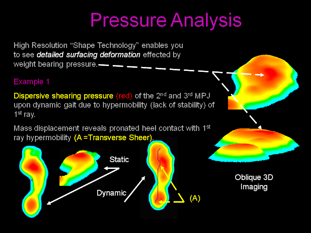
Why is 3DO Imaging Superior to Casting -  Orthotics are manufactured based on a 60 year old "Art Form" and therefor is full of errors.   3DO Imaging evaluates Static and Dynamic body mechanics (Mass Displacement Analysis - Motion Analysis - Pressure Analysis - Body Balancing Analysis - Symmetry - Gait Analysis - 3D Geometry) where Neutral Joint Positioning is computed by Software for accurate joint alignment and function.  Our focus is the total body, not just the foot.   

Testing - We provide $750.00 worth of analytical testing report for free.   
  1. 3DO Weight Bearing Kinematic Imaging Analysis (Static Non Movement) and (Dynamic Movement).    A outcome measurement report will be provided to you.
  2. High Resolution Optical Imaging
  3. Video Gait Analysis  


 Testing and examination time:   15-20 minutes
 
Best clothing to wear
  • Shorts
  • Pants or clothing that we can roll up to the lower leg for proper testing.
Manufacturing Time:    48 to 72 hours for Custom Bio Engineered Devices and 10 days for Custom Hand Made Ergonomic Footwear.


Locations: By Appointment Only (Monday - Friday) - Saturdays by Special Appointment
  • Digital Orthotics Inc., Technical Support Center - 222 Fashion Lane, Suite 107 - Tustin CA 92780 (714) 669-9600.  The map below provides directions from our old office to our new location.
 

  • Digital Orthotics Inc., Riverside Production Unit - 3293 Trade Center Drive - Riverside CA 92507 (951) 686-2600 - Testing and Analysis performed by John Giraldo, Production Lab Manager.


Thursday, November 15, 2012

Public Blog Introduction

Public - Non Physician Blog Site

This blog site provides a variety of important articles on injuries, disease and prevention.   Feel free to share these blogs with your friends and family.


Tuesday, November 13, 2012

Childrens foot and limb diseases

Pediatric (Children's) Foot & Limb Diseases
 
 
Pediatric Foot and Limb Diseases are easily treatable.   "The earlier, the better", States Dr Craig Lowe, (Researcher).   As bones are soft and pliable, Orthotics can be used to assist in proper development of the bones of the feet. 
 
Bad Press. . .
Unfortunately, Pediatricians receive little training regarding Orthopedic diseases in their Residency.   When parents take the child to the doctor, they pass off concerns as, "don't worry, they will grow out of it". . . "They don't grow out of these diseases.   We see them as surgical patients in their 20's and 30's", stated Dr Lowe.    Unless the deformities are substantial, they usually do not refer to either Pediatric Orthopedists or Podopediatric Specialists.
 
Early Diagnosis and Treatment is the Key
Unless deformities are severe, start evaluations at age 5 after they start walking.  
  • Note the age they started walking.  Generally we will see children beginning ambulation at the age of 1 year old and crawling at 9 months.
  • 3DO Imaging should be done starting at age 5 and repeated yearly as they grow.
  • Most Pes Planus Splayed Flatfoot looking feet usually reduce by age 5 which is when you can start treatment.
Types of Common Deformities After the Age of 5
 
Gait
  • Adducted - "Intoe Gait"
  • Abducted - "Outtoe Gait"

Foot
  • Metatarsus Adductus - "C" Shaped Foot
  • Pes Planus
  • Club Foot
Knee
  • Medial Knee Position
Hip
  • Dislocating Hip
  • External Hip Position
  • Internal Hip Position


    Diabetic Footwear

     
    Custom Diabetic Footwear
     
    Type 1 (Insulin Dependent) versus Type 2 (Adult Onset) Diabetes
     
    Type 1
    They are currently taking Insulin to control blood sugar
    They are usually on a strict diet
    They may or may not have peripheral neuropathy
    Thay may have Charcot Joint Disease as well
    Manditory 3DO Imaging (3D Weight Bearing Kinematic Imaging), to evaluate static and dynamic linear and sheer pressure loading of the feet.   Optical High Resolution Imaging for Plantar Lesions.
    • This needs to be done every 6 months with open lesions (ulcers) and yearly with no ulcerations and every 12 months with no ulcerations.
    • Use 3DO Imaging to demonstrate migratory neuropathic pressure variants which change as disease changes.
    • Predict future ulceration sites.
     
    Type 2
    They are on Diet and Oral Hypoglycemics
    Usually there is no peripheral neuropathy 
    Manditory 3DO Imaging to evaluate static and dynamic linear and sheer pressure loading of the feet.   Optical High Resolution Imaging for Plantar Lesions.
     
    The enemy. . . Sheer Pressure. . .
    Sheer pressure creates transverse plane "slide" of metatarsal heads during midstance.   More so in the hypermobile foot.  Check for equinus conditions which will increase plantar loading.


    The Solution. . . Custom Diabetic Orthotics


    Flex Polypropylene Shell + Poron and Plastizote Top Cover


    How to make fashion footwear work better


    How to make Fashion Footwear Work Better

     
    Yikes !  Fashion Pumps and Gorgeous Footwear
     
    How we men love them. . .
     
    Lets face it, fashion will always drive women to look their best.   Women who are fashion orientated will never get rid of their pumps.
     
    What we have done is enable the foot to better function in high fashion shoes.   This is done through the "Style Orthotic" which is designed to enhance biomechanical function in extreme fashion shoes.
     
    The problems of Orthotics and fashion footwear are (2);
    1. Bulkiness of the custom device - It needs to be a low profile custom Orthotic.
    2. Integration of the custom device into the Fashion Shoe - We need to have the shoe so the Orthopedic Technician can properly integrate the device for a perfect fit, (Footwear Integration). 
    The symptoms should be obvious;
    • Increased metatarsal head pressures due to rearfoot to forefoot position as the ankle joint is plantarflexed.
    • Metatarsal Neuromas - Metatarsalgia - Plantar Metatarsal Tylomas and Callouses.
    • Hammertoes - Mallettoes - Bunions (1st and 5th MPJ)
    • Shin Splints
    • Lumbar Spinal Pain due to extenuated Lordosis
    • Aggravates Equinus conditions 
    Treatment:
    • 3DO Imaging (Static and Dynamic) for 3D imaging and analysis of peak load pressures
    • Style Orthotics with Poron Sulcus Extension
    • Optional Metatarsal Rise to further offload peak MPJ pressures
    • Metatarsal head pockets to allow the metatarsal heads to offload better.
    • Good Midtarsal Joint Control